BRAVO
DR
VMT
ERM
Características principales
Herramientas esenciales para su éxito
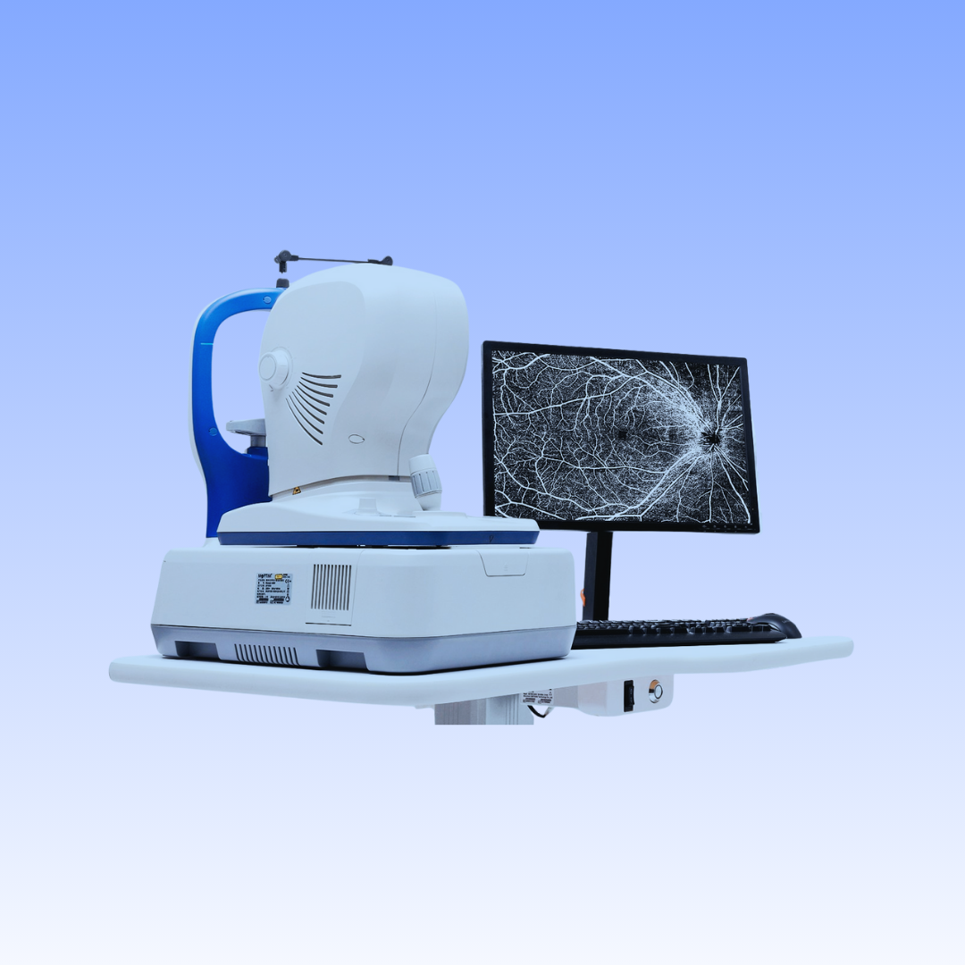
Real-time SLO with eye tracking
Simultaneously acquires OCT images and 45 degrees fundus images based on Scanning Laser Ophthalmoscope (SLO), providing a real-time overview of the retina that allows easy localization of the lesion area before acquisition.
To minimize the artifacts caused by eye drift and micro saccades, it uses SLO-based eye tracker, which gives you more confidence in practice.
16mm angle-to-angle analysis
16mm angle-to-angle anterior scan with data analysis.
Deep Choroidal Imaging (DCI) mode
Using Deep Choroidal Imaging for detection of choroidal neovascularization.
Comprehensive software analysis
provides 9 scan patterns to help you improve diagnostic efficiency:
- Retina (HD line, Six-Radial lines, Multi, 3D Cube 10x10mm)
- Glaucoma (Glaucoma Disc for ONH analysis, Glaucoma Macular for GCC analysis)
- Cornea (HD line, Six-Radial lines, Angle-to-Angle)
The software analysis features are always up-to-date and free for upgrade (excluding OCTA module).
Ficha Tecnico
Todo lo que necesita para comenzar.
OCT IMAGING
Methodology Spectral domain OCT
Optical source Super luminescent diode (SLD), 840 nm
Scan speed 50,000 A-scans/s
Axial resolution (optical) 5 microns (optical), 2.7 microns (digital)
Transverse resolution 15 microns (optical), 3 microns (digital)
A-scan depth 3.1mm
Diopter range –20 a +20 diopters
Working distance 30 mm
Scan patterns Macular: HD line scan (6 / 12 mm), 3D scan (10 mm x 10 mm), 6-line radial scan, Multi (X-Y: 5 x 5, X:9, Y:9) Disc: 3D scan (6 mm x 6 mm)
Disco óptico: Exploración 3D (6 mm x 6 mm)
Anterior: HD line scan (6 / 16mm), 6-line radial scan, angle-to-angle scan (16mm)
FUNDUS IMAGING
Methodology Line scanning laser ophthalmoscopy (LSLO)
Minimum pupil diameter 3.0 mm
Field of view 45 ± 1 degrees
ELECTRICAL AND PHYSICAL
Weight 30.5 kg
Dimension 532 mm (L) x 360 mm (W) x 540 mm (H)
Source voltage AC 100 – 240 V
Frequency 50 Hz – 60 Hz
Power input 90 VA